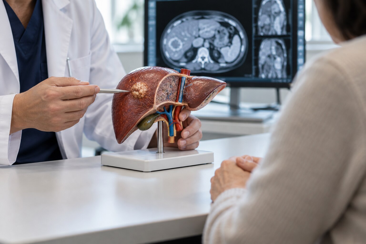
Surgery is considered when doctors can remove all visible tumor while preserving enough functioning liver and maintaining blood flow and bile drainage. By the end, you will know which diagnoses prompt this discussion, how scans and liver-reserve tests determine feasibility, which alternatives may be safer, and what recovery and follow-up involve.
Key takeaways
- Removal is considered when the tumor can be cleared while preserving enough working liver.
- CT or MRI scans map tumor location, blood vessels, bile ducts, and spread.
- Liver function tests and background liver disease determine surgical safety.
- A multidisciplinary team compares resection, ablation, embolization, and systemic treatment.
Which liver tumor diagnoses lead doctors to discuss removal?
Removal enters serious discussion when the diagnosis is confined to the liver or liver-dominant disease can be completely cleared while preserving enough functioning tissue. The diagnosis matters, but location, spread, liver health and tumor biology decide whether removal is realistic.
| Diagnosis | Why removal may be discussed | What “advanced” may mean |
|---|---|---|
| Hepatocellular carcinoma | A removable tumor in a well-functioning liver, especially without major portal hypertension | Large size, multiple tumors, vascular involvement or cirrhosis; advanced does not automatically mean inoperable |
| Intrahepatic cholangiocarcinoma | A localized bile-duct cancer that can be removed with adequate margins and reconstruction | Involvement of major vessels, bile ducts or nearby organs |
| Colorectal liver metastases | Liver-limited or liver-dominant spread that can be completely cleared, even with a narrow negative margin | Several tumors, difficult locations or disease requiring chemotherapy before surgery |
| Neuroendocrine metastases | Slow-growing, liver-dominant metastases when complete or useful tumor reduction is achievable | Multifocal disease or spread requiring staged treatment or other liver-directed therapy |
| Selected benign tumors | Hepatocellular adenoma or a symptomatic hemangioma causing bleeding risk, pain or uncertainty | Size, symptoms, growth or uncertain diagnosis rather than cancer spread |
“Advanced” describes extent or technical difficulty, not an automatic verdict against surgery. Surgeons ask whether they can remove all visible tumor while preserving adequate inflow, outflow, bile drainage and liver reserve. Cirrhosis, jaundice, ascites, low albumin or poor general health can make removal unsafe because postoperative liver failure becomes the greater danger.
How do scans and liver function establish whether surgery is feasible?
Feasibility depends on removing all visible tumor while leaving enough well-functioning liver with intact blood flow and bile drainage. A large solitary tumor can be removable, while a smaller tumor may be unsafe if it involves major vessels, bile ducts, or too much of the remaining liver.
Doctors assess the whole liver and the chest, abdomen, and pelvis rather than measuring the largest mass alone:
- Multiphasic contrast CT shows tumor enhancement, number, location, vascular invasion, and possible spread outside the liver.
- Liver-protocol MRI gives detailed views of small lesions, bile ducts, and the boundary between tumor and normal liver.
- Ultrasound assesses blood flow through the portal and hepatic veins and supports image-guided evaluation.
- PET-CT for selected metastatic disease helps identify cancer deposits outside the liver that would change the treatment plan.
Volumetry calculates how much liver would remain after resection. Surgeons also review whether the remaining tissue will retain adequate portal inflow, hepatic venous outflow, and biliary drainage. A planned remnant that is too small or poorly supplied creates a risk of post-hepatectomy liver failure.
Blood tests measure bilirubin, albumin, clotting, and platelet levels. Doctors also consider cirrhosis and clinically significant portal hypertension; uncertain liver reserve may require functional testing. Chemotherapy can further reduce reserve through oxaliplatin-related sinusoidal injury or irinotecan-related steatohepatitis, so its timing matters.
Which operation or alternative treatment best fits the tumor?
The best treatment balances complete tumor control against the liver left behind. Surgeons choose a wedge resection for a small, peripheral lesion; a segmentectomy for disease confined to one anatomical segment; a sectionectomy for tumors spanning several adjoining segments; and a hemihepatectomy when one side of the liver must be removed.
Location near the hepatic veins, portal vein, bile ducts, or major arteries can require vascular reconstruction or make another treatment safer.
| Option | What it means | When it applies |
|---|---|---|
| Resection | Removes visible tumor with a margin while preserving functioning liver | Preferred for technically removable disease, adequate liver reserve, and a realistic chance of complete control |
| Ablation | Destroys a tumor with heat or another needle-based energy | Useful for small, favorably located tumors when surgery would sacrifice too much liver |
| Transarterial treatment | Delivers chemotherapy or radiation through tumor-feeding arteries | Used for selected liver-confined tumors that are unresectable or need bridging or downstaging |
| Systemic therapy | Treats cancer throughout the body with targeted drugs or chemotherapy | Favored for widespread, biologically aggressive, or unresectable metastatic disease |
| Immunotherapy | Activates immune responses against cancer | Used for cancers with approved immunotherapy indications, alone or with other systemic drugs |
| Transplantation | Replaces the diseased liver and removes the tumor-bearing organ | Considered for selected HCC patients, including those within Milan criteria or successfully downstaged |
A multidisciplinary team may change the plan after systemic therapy, portal-vein embolization, ablation, or transplant assessment. For metastatic disease, preoperative chemotherapy can reveal tumor behavior, but oxaliplatin-related sinusoidal injury and irinotecan-related steatohepatitis can reduce surgical liver reserve.
What happens during the multidisciplinary evaluation before consent?
The recommendation comes from a multidisciplinary tumor board, not from scan size alone. The hepatobiliary surgeon, liver radiologist, medical oncologist, radiation specialist, anaesthetist and, when relevant, transplant specialist review the diagnosis, biopsy or resection pathology, multiphasic CT or liver-protocol MRI, chest imaging, PET-CT findings, blood tests, cirrhosis status, prior chemotherapy and other illnesses.
They ask whether all visible disease can be controlled while preserving adequate blood flow, bile drainage and functioning liver.
Before consent, request clear answers to these questions:
- What exactly will be removed, and where are the tumor, hepatic veins, portal vein and bile ducts in relation to the planned cut?
- What is the predicted future liver remnant, and how was its function assessed rather than estimated from volume alone?
- Do I need portal vein embolization to enlarge the future remnant, and what is the risk of tumor progression during the waiting period?
- If disease is spread across both lobes, would a two-stage hepatectomy be safer than one operation, and why is ALPPS being considered or rejected?
- How have chemotherapy-related injuries, including oxaliplatin-associated sinusoidal damage or irinotecan-associated steatohepatitis, affected timing?
- What alternatives offer comparable control if resection is unsafe?
Ask Dr Mayur Porwal 08042753618 to explain which specialists reviewed your case, the intended margin, possible need for transfusion or drainage, and what finding would stop the operation after it begins. Complete nutrition, anaesthesia and medication reviews before signing consent.
Related services
Liver (Tumors) and Infection The liver can be affected by tumors (benign or malignant) and infections, both of which can significantly impact liver function and overall health. View service → |
Abdominal Tumor Surgery An abdominal tumor is an abnormal growth of tissue in the abdominal cavity. View service → |
What risks, recovery, pathology, and follow-up should patients expect?
The central trade-off is control versus loss of functioning liver. A margin-negative resection lowers the chance of leaving tumor behind, but removing more tissue can reduce the future liver remnant and increase the risk of liver failure, especially with cirrhosis, jaundice, low albumin, or portal hypertension.
- Bleeding can require transfusion or another procedure.
- A bile leak can prolong drainage and hospital care.
- Infection can delay recovery or require antibiotics and drainage.
- Liver failure can cause jaundice, fluid accumulation, confusion, and intensive-care treatment.
Expect hospital recovery followed by several weeks of reduced stamina, restricted lifting, and gradual return to normal eating and activity. The pathology report confirms the tumor type, microscopic margin status, grade, and invasion pattern; these findings determine whether the operation achieved complete clearance and whether additional treatment is warranted.
For colorectal metastases, a margin under 1 cm can still be acceptable when microscopic clearance is complete.
Follow-up combines scheduled liver imaging with blood tests. Doctors may track alpha-fetoprotein and carcinoembryonic antigen when those markers apply to your tumor, then adjust surveillance after treatment response, pathology risk, and liver function are known. Recurrence can lead to repeat surgery, ablation, transarterial treatment, systemic therapy, immunotherapy, or transplant evaluation rather than automatic reoperation.
Frequently asked questions
Which liver tumor diagnoses can lead doctors to discuss removal?
Doctors discuss removal for tumors confined to the liver and selected liver-dominant cancers that can be completely cleared while preserving adequate functioning liver tissue. The diagnosis, tumor number and size, spread, and tumor biology all affect feasibility.
How do scans and liver function establish whether surgery is feasible?
Contrast-enhanced CT or MRI maps tumor size, number, location, blood-vessel involvement, bile-duct involvement, and spread. Blood tests and liver assessment examine bilirubin, albumin, clotting, platelet count, and the effects of cirrhosis or other liver disease.
How do doctors choose between liver resection and other treatments?
The team matches treatment to tumor location, size, number, liver reserve, spread, and overall health. Options can include liver resection, ablation, embolization, transplantation, systemic treatment, or a combination.
What happens during multidisciplinary evaluation before consent?
Surgeons, hepatologists, oncologists, radiologists, pathologists, and anaesthetists review the diagnosis, scans, liver reserve, surgical plan, alternatives, and expected risks before consent.
What should patients expect after liver tumor removal surgery?
Expect hospital monitoring, pain control, gradual return to eating and movement, and follow-up for complications such as bleeding, infection, bile leakage, liver failure, or blood clots. Pathology confirms the tumor type and margins, while follow-up scans and blood tests check for recurrence.
Related service
Liver surgery involves the removal or treatment of diseased parts of the liver due to tumors, cysts, infections, trauma, or other liver conditions. View service → |